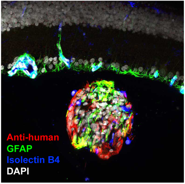
Figure 4.
Intravitreal graft of retinal progenitor cells. Cultured human RPCs, labeled with antihuman antibody (red), are seen following injection into the vitreous cavity of a rat eye. The cells are injected as a single cell suspension but subsequently aggregate in vivo to form small clusters, as seen here. These clusters are free-floating and provide neutrotrophic support to the retina (laminar structure above the graft) without the need for integration into the host tissue. The RPCs of the grafts differentiate along either neuronal or glial lineages, with the latter seen here by way of labeling for GFAP (green). Host mononuclear leukocytes investigate the donor cells, illustrated here by positivity for isolectin B4 (blue) within the graft, but do not elicit an immunological rejection response. Nuclei are labeled with DAPI (white). This image was provided by Dr. Geoffrey Lewis, UCSB.

