
Figure 9.
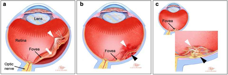
Schematic diagram depicting selected complications of retinal cell therapy. (a) Retinal detachment and proliferative vitreoretinopathy. The retina has detached (white arrow) and the detachment involves the fovea, thus severely reducing visual acuity. The detachment depicted here is associated with a proliferative vitreoretinopathy membrane (white arrowhead) and a retinal break (clear arrowhead). (b) Hemorrhage. Here, blood has collected in the vitreous cavity and on the retinal surface (vitreous cavity hemorrhage, white arrowhead) and beneath the retina (subretinal hemorrhage, black arrowhead) causing a hemorrhagic retinal detachment that threatens the fovea. (c) Epiretinal membrane. A thick epiretinal membrane (white arrowhead) has resulted in foveal thickening and distortion (black arrowhead). Epiretinal membranes can form after cell delivery regardless of whether the exogenous cells have been inadvertently placed in the epiretinal surface. with or without Complications that are not depicted in this diagram include infection (endophthalmitis), cataract, elevation of intraocular pressure and crystalline lens dislocation. (Illustration by Timothy Phelps, MS, FAMI.)