Fig. 1.
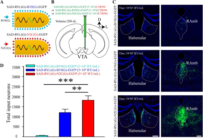
The retrograde gene transduction efficiency of SAD-RV(ΔG)-N2C(G)-EGFP is higher than that of SAD-RV(ΔG)-B19(G)-EGFP. A Schematics of the virion structures of SAD-RV(ΔG)-B19(G)-EGFP (upper panel) and SAD-RV(ΔG)-N2C(G)-EGFP (lower panel). B Schematic of the in vivo tracing study. A low dose of CTB594 was co-injected with either a low titer of SAD-RV(ΔG)-B19(G)-EGFP (5 × 10⁷ infectious units (IFU)/mL), a high titer of SAD-RV(ΔG)-B19(G)-EGFP (5 × 10⁸ IFU/mL), or SAD-RV(ΔG)-N2C(G)-EGFP (5 × 10⁷ IFU/mL) into the VTA of C57 mice. C Many regions upstream of the VTA, such as the habenular nucleus (left panels) and the midbrain raphe nuclei (RAmb, right panels), were retrogradely labeled by SAD-RV(ΔG)-N2C(G)-EGFP (lower panels) or SAD-RV(ΔG)-B19(G)-EGFP with different titers (upper and middle panels). D Numbers of neurons in the whole brain retrogradely infected by a low (5 × 10⁷ IFU/mL) or a high (5 × 10⁸ IFU/mL) titer of SAD-RV(ΔG)-B19(G)-EGFP, or SAD-RV(ΔG)-N2C(G)-EGFP (5 × 10⁷ IFU/mL). Scale bars, 200 µm; n = 4 mice/group; *P < 0.05, **P < 0.01, ***P < 0.001. n.s., no significant difference; one-way ANOVA followed by the LSD multiple comparison test. The nuclei were stained blue by DAPI.

