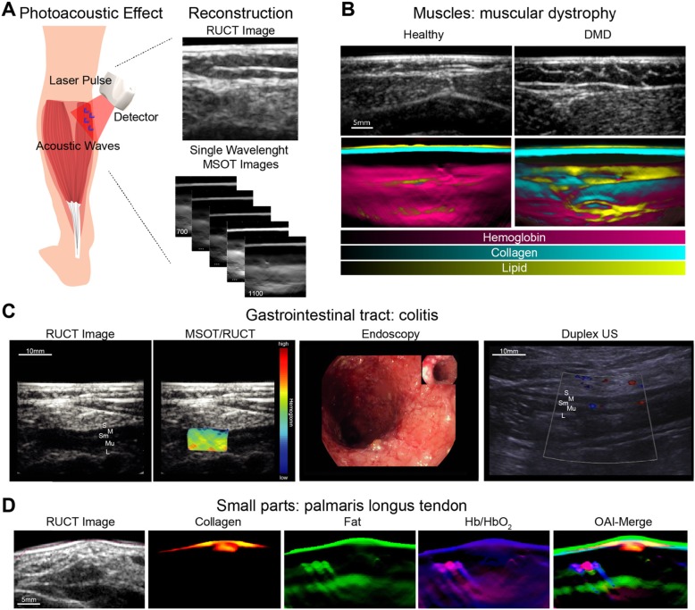
Fig. 1.
Principle of optoacoustic imaging. a The terms “photoacoustic” or “optoacoustic” describe the use of “light” and “sound.” The illumination of tissue by pulsed laser light and the subsequent recording of thermoelastic pressure waves combines the advantages optical (high contrast) and acoustic (high resolution) imaging. Subsequent image reconstruction and spectral unmixing enables non-invasive real-time visualization and quantification of specific endogenous chromophores such as hemoglobin, lipids, and collagens. RUCT reflectance ultrasound computed tomography, MSOT multispectral optoacoustic tomography. b Imaging example from muscular imaging resolving hemoglobin, collagen, and fat signals. DMD Duchenne muscular dystrophy. c MSOT imaging of the colon unmixed total hemoglobin signal. S serosa, M muscularis mucosa, Sm submucosa, L lumen, US ultrasound. d Example for imaging of palmaris longus tendon at the wrist with unmixing for different tissue components. Note the clear delineation of yellow/red containing tendon in comparison of hemoglobin signals from vessels

