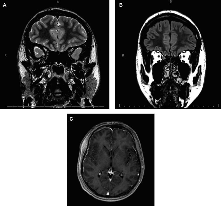
FIGURE 7.
Case 3. Preoperative MRI. A, Oblique coronal T2W image shows indistinct cortex in the left frontal region with T2 prolongation extending deep into the white matter. B, Coronal T2W FLAIR demonstrates the same left frontal abnormality. C, Three-dimension magnetization-prepared rapid gradient echo T1W axial image with gadolinium.

