Figure 3.

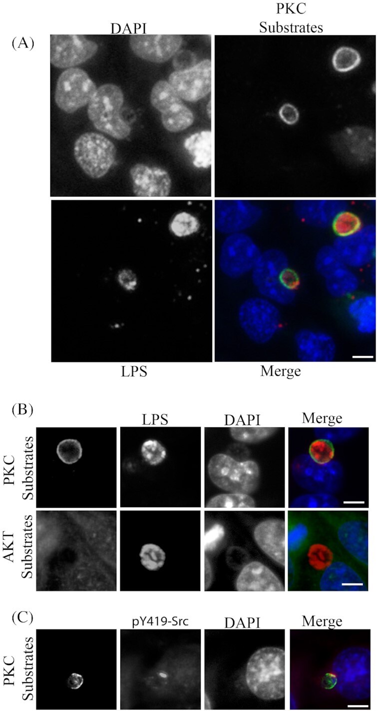
PKC phosphorylated substrates are recruited to the entire periphery of the C. trachomatis inclusion. HeLa cells were infected with C. trachomatis L2 at an MOI of 0.5 for 18 hours, fixed in cold fixative and processed for immunofluorescence microscopy. (A) Phospho (Ser)-PKC substrates are shown surrounding the chlamydial inclusion (Chlamydia detected with anti-Chlamydia LPS). Multiple infected and uninfected cells can be seen for comparison. (B) Phospho (Ser)-PKC substrates and Akt substrates were detected with phosphospecific antibodies for recruitment to the chlamydial inclusion (Chlamydia detected with anti-Chlamydia LPS). (C) Active Src kinases (pY49-Src) are shown colocalizing in discrete microdomain overlapping the phospho-(Ser)-PKC. Scale bar, 10 µm.
