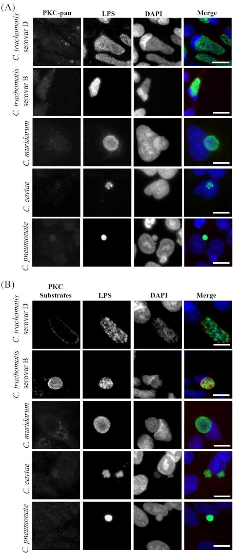
Figure 4.

PKC and PKC substrate recruitment are limited to C. trachomatis serovars. HeLa cell monolayers were infected with C. trachomatis serovar D (42 hours), C. trachomatis serovar B, (42 hours), C. muridarum (18 hours), C. caviae (18 hours) and C. pneumoniae (42 hours) at an MOI of ∼0.5, fixed in cold methanol and processed for immunofluorescence microscopy. (A) Total phospho-PKC recruitment as detected by a phospho-PKC-pan antibody (arrows indicate discrete regions of phospho-PKC recruitment) and (B) recruitment of phosphorylated PKC substrates are shown. All Chlamydia species were detected with anti-Chlamydia LPS antibody. Scale bar, 10 µm.
