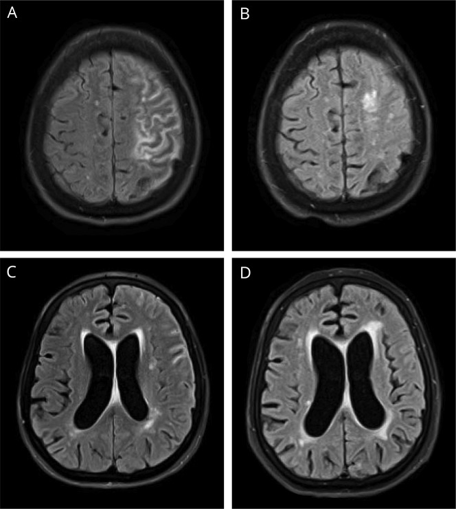
Figure 1. FLAIR MRI.
Noncontrast FLAIR MRI of the head, axial section. For interval comparison, images A and C are from November 11, 2016, and (B and D) from April 18, 2017. (A) Sulcal hyperintense signal compatible with meningitis with mild amount of hyperintense signal in subcortical white matter of the frontal and parietal lobes suggesting inflammation or infection. (B) Significantly decreased sulcal hyperintense signal in the left cerebral hemisphere with mild amount of remaining hyperintensity in superior portions of the left frontal and parietal lobes. (C) Sulcal hyperintense signal with mildly enlarged lateral ventricles. (D) Sulcal hyperintense signal is no longer seen, but still with mildly enlarged lateral ventricles. There is mildly increased amount of periventricular white matter FLAIR hyperintense signal.

