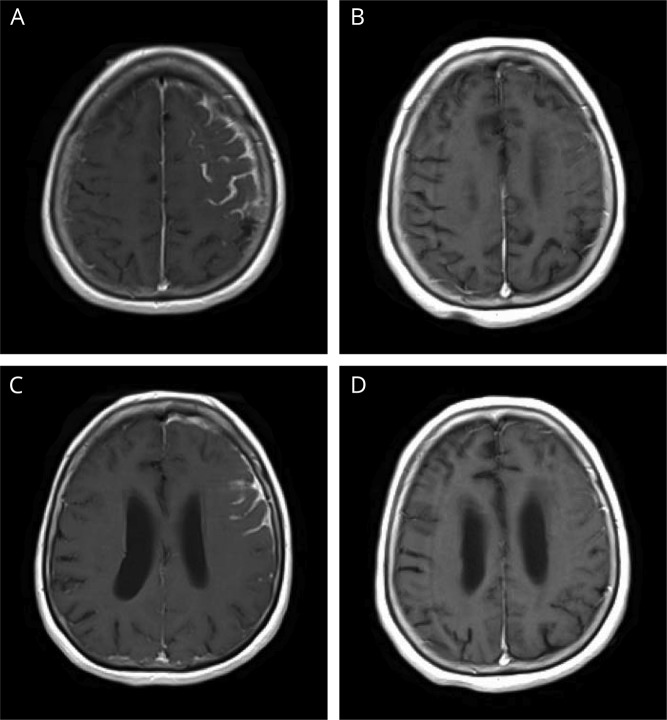
Figure 2. Contrast T1-weighted MRI.
Contrast T1-weighted MRI of the head, axial section. For interval comparison, images A and C are from November 11, 2016, and (B and D) from April 18, 2017. (A and C) Leptomeningeal and pachymeningeal enhancement are seen in the left frontal lobe. (B) Resolution of previous enhancement with continued mild sulcal effacement in the left frontal and parietal lobes. (D) Minimal remaining leptomeningeal enhancement when compared with prior.

