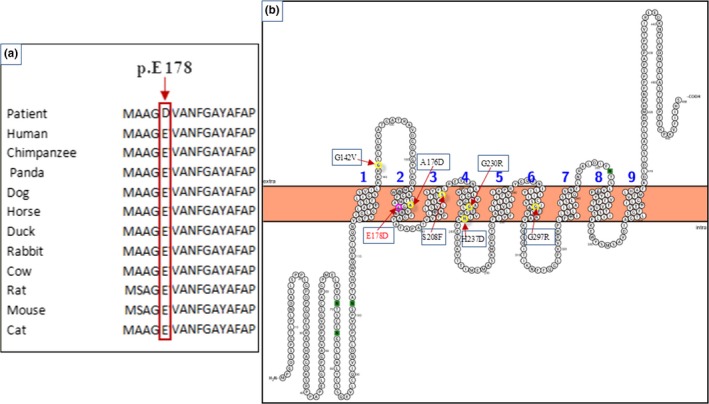
Figure 2.

(a): Sequence alignment of the NIPA4 protein in different species performed by the Clustal OMEGA program and showing the conservation of the Glutamic acid residue at position 178 throughout species. (b): Predicted transmembrane structure of the NIPA4 protein performed by Protter program
