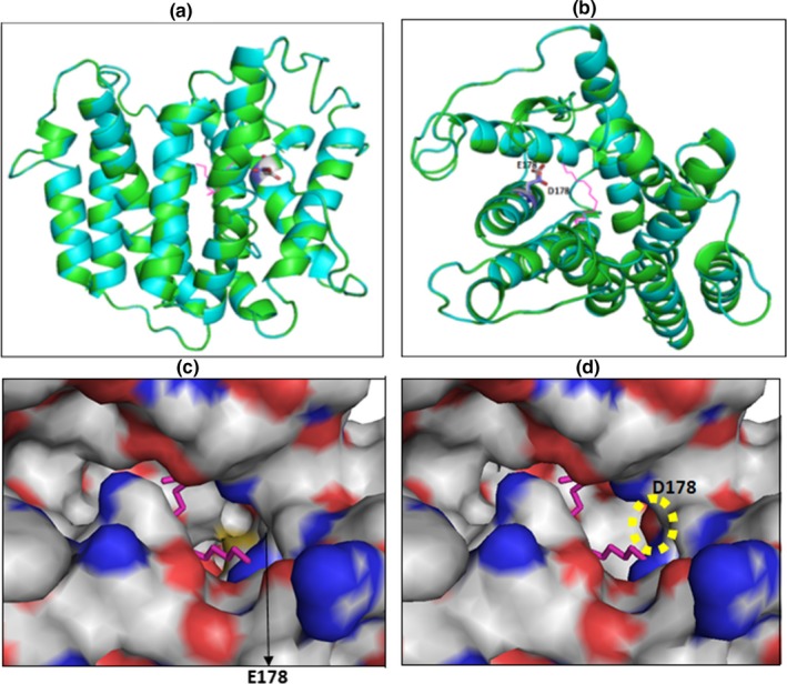
Figure 3.

Superimposition of the Nipa4 wild type and mutant model structures. (a): Ribbon representation of the superimposed models, displaying residue 178 (Glu in wildtype and Asp in the mutant) as sticks. To localize the potential transport channel of Nipa4, the original channel‐bound ligand from the template is shown as magenta sticks. (b): upper view from (a) showing clearly the potential transport channel. (c), Zoom surface representation (from b) showing residue 178 in the transport channel of wild type Nipa4. (d), Zoom surface representation (from b) showing residue Asp178 in the transport channel of the Nipa4 mutant. The negatively charged oxygen of Asp178 carboxyl group located in the transport channel is surrounded by an open circle. In c and d panels, oxygen and nitrogen are colored in red and blue, respectively
