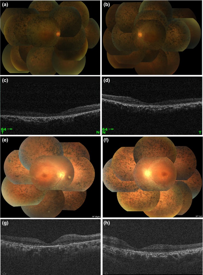
Figure 2.

Color fundus puzzle and spectral‐domain optical coherence tomography (SD‐OCT) of macular regions of two probands. (a, b, e, and f) Color fundus puzzle of the proband II‐2 (family No.1: ARRP‐01) and the proband II‐1 (family No.2: ARRP‐02) bilaterally show the typical symptoms of RP, characterized by optic disc waxy pallor, attenuated retinal vessels, and the retina is atrophied and the color is blue‐gray. (c, d, g, and h) SD‐OCT of the macular of the probands II‐2 (ARRP‐01) and II‐1 (ARRP‐02) show the degenerative changes of retinal layers in both eyes, revealing the structural damages of both inner segment ellipsoid band and photoreceptor outer segment
