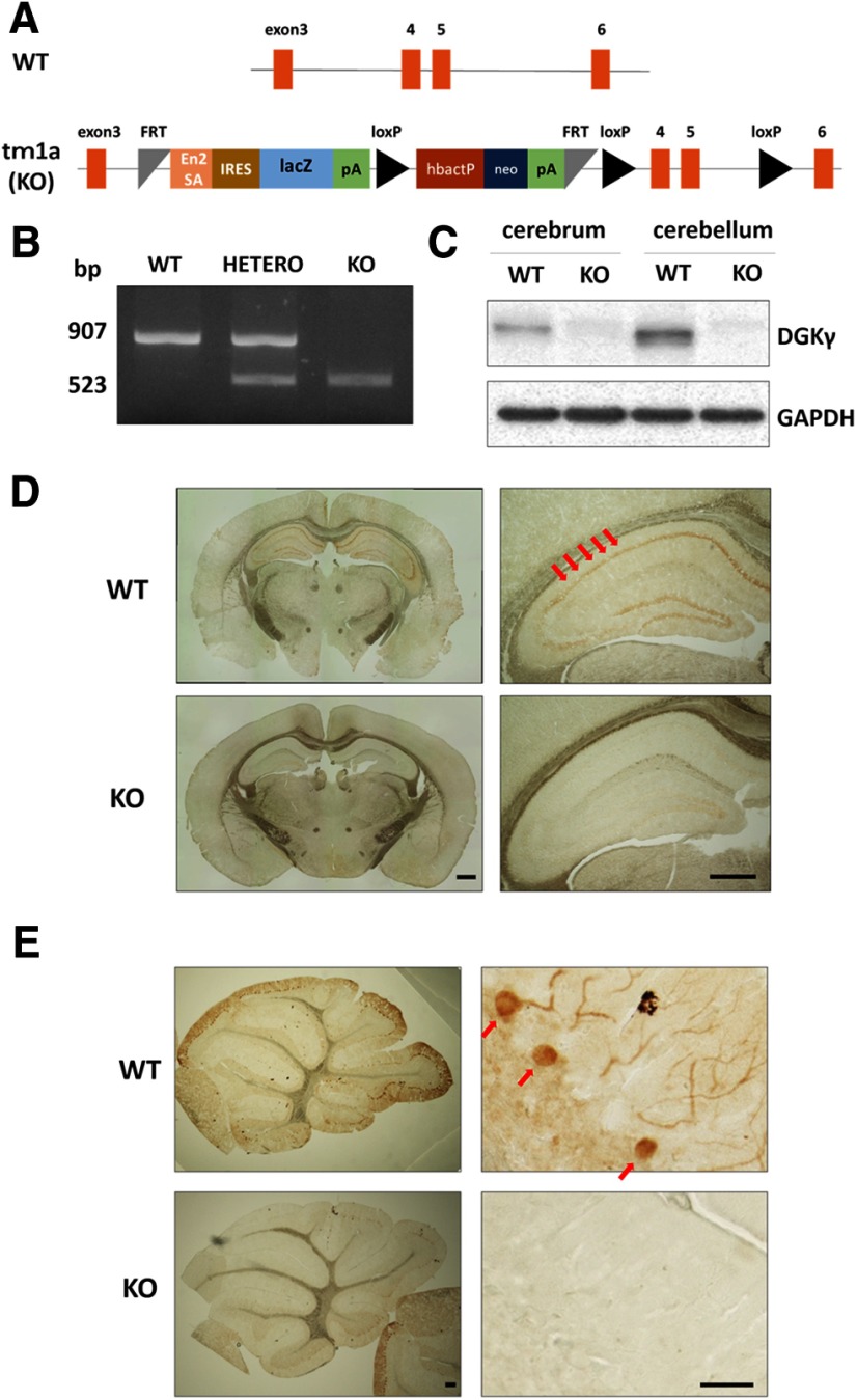
Figure 1.
Generation of DGKγ KO mice and PCR genotyping. A, Promotor-driven cassette inserted between exons 3 and 4 of the gene that encodes DGKγ. En2SA, engrailed 2 splice acceptor; lacZ, β-galactosidase; pA, adenovirus polyadenylation signal; loxP, Cre recombinase recognition sequence; hbactP, human-β-actin promotor; neo, neomycin. B, Typical result of PCR genotyping. Bands at 907 and 523 bp were expected for the WT and tm1a alleles, respectively. C, Cerebral and cerebellar lysates from WT and DGKγ KO mice were subjected to Western blotting and probed with an anti-DGKγ antibody. D, E, Coronal sections of the cerebrum (D) and parasagittal sections of the cerebellum (E) were subjected to immunohistochemistry and stained with an anti-DGKγ antibody. The red arrows show the hippocampus and Purkinje cells. Scale bars: 500 μm (D, right and left, E, left) and 100 μm (E, right).

