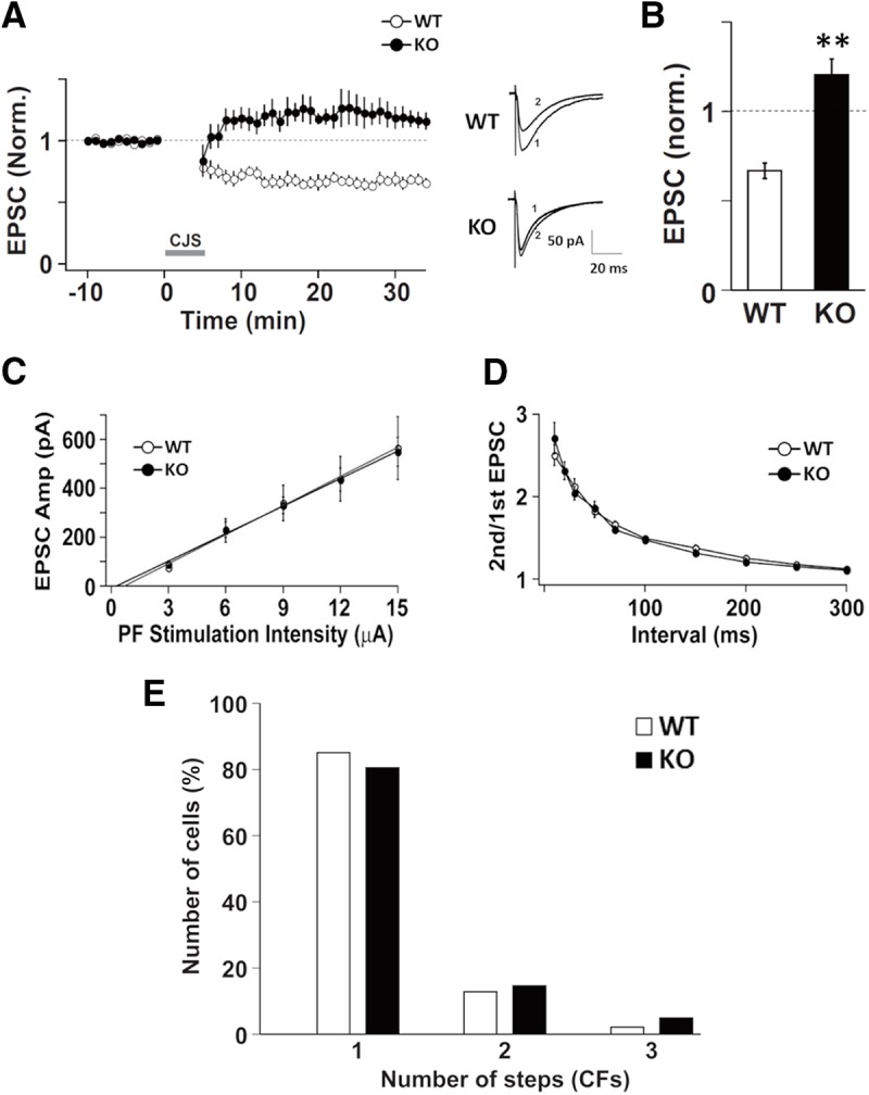
Figure 3.

Electrophysiological properties of Purkinje cells in DGKγ KO mice. A, Changes in the PF-EPSC amplitude before and after the CJS of PFs (1 Hz, 300 s) with depolarization applied at time 0. The PF-EPSC amplitude was normalized to the mean over 10 min before CJS (WT: n = 5; KO: n = 5). Sample traces immediately before (1) and 30 min after (2) CJS. B, Average PF-EPSC amplitude over the 21- to 30-min period after CJS; **p < 0.01, followed by Student’s t test. C, The input-output relationship of PF-EPSCs. The PF-EPSCs amplitudes of Purkinje cells from WT and DGKγ KO mice plotted as a function of stimulus intensity (WT: n = 11; KO: n = 10). D, The PPR of PF-EPSCs recorded from Purkinje cells from WT or DGKγ KO mice at several interpulse intervals (WT: n = 11; KO: n = 10). E, Summary histograms showing the percentage of discrete steps of CF-EPSCs (WT: n = 47; KO: n = 41). Data are expressed as mean ± SEM.
