Figure 1.
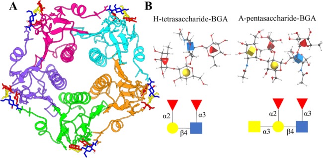
(A) Crystal structure of the pentameric cholera toxin B subunit in complex with glycans. Each protein chain is shown in a different color, and the glycan is represented in the ball-and-stick model. (B) Structure and nomenclature of Lewis Y (LeY) blood group determinants: H-tetrasaccharide and A-pentasaccharide BGAs. The structure and nomenclature of LeY blood group determinants are displayed in three-dimensional (3D) symbol nomenclature for glycans (SNFG) icon mode. In this representation, N-acetylglucosamine is shown as a blue cube, l-fucose is shown as a red cone, d-galactose is shown as a yellow sphere, and N-acetylgalactosamine is shown as a yellow cube. All images were generated using Visual Molecular Dynamics (VMD) with the help of the 3D-SNFG plugin available at www.glycam.org/3d-snfg. A detailed atomic structure is also shown in the ball-and-stick model, where the standard color scheme is used to depict different types of atoms.

