Figure 6.
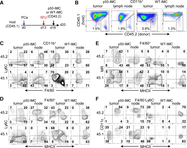
p50-IMC generates increased PCa tumor and lymph node myeloid cells and dendritic cells compared with WT-IMC. (A) CD45.1+ mice inoculated with PCa on day 0 received 5FU on day 13 and 107 p50-IMC or WT-IMC derived from CD45.2+ mice on day 18, followed by tumor and inguinal lymph node mononuclear cell isolation and analysis on day 20, as diagrammed. Data acquired from 2×10⁴ CD11b+ cells per mouse from five mice in a single experiment were pooled for analysis. (B) CD11b+ myeloid cells were analyzed for CD45.2+ (p50-IMC donor-derived) and CD45.1+ (host) cells by flow cytometry. (C) IMC and host myeloid cells were analyzed for Ly6C and F4/80. (D) F4/80+ myeloid cells within the CD45.2+ and CD45.1+ populations were analyzed for Ly6C and MHCII. (E) F4/80+ myeloid cells within the CD45.2+ and CD45.1+ populations were analyzed for MR and MHCII. (F) F4/80−Ly6C− myeloid cells within the CD45.2+ and CD45.1+ populations were analyzed for CD11c and MHCII. 5FU, 5-fluorouracil; IMC, immature myeloid cell; MHCII, Major Histocompatibility Complex II; MR, magnetic resonance; p50-IMC, p50-negative immature myeloid cell; PCa, prostate cancer; WT, wild type.

