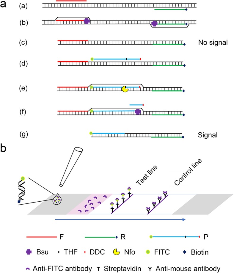
Fig. 1.

Schematic diagram of the RPA-LFD method. (a) Principle of RPA amplification. DNA strands are presented as horizontal lines, and base pairings are indicated as short vertical lines between the DNA strands. The forward primer (F), reverse primer (R), probe (P), Nfo, Bsu and modifications on DNA are indicated with different shapes and colors. The legends are given at the bottom of the image. (b) Schematic diagram of the working principle of the lateral flow dipstick. The sample pad is indicated by the gray parallelogram on the left, the absorbent pad is indicated by the gray parallelogram on the right, and the conjugate pad is shown in pink. Liquid migration direction is indicated by an arrow. Molecules could be trapped by the materials on the test line, and the control line is indicated by different shapes. Shapes and their representative molecules are listed at the bottom of the image
