Fig. 1.

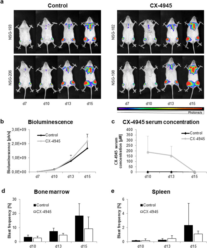
Evaluation of CX-4945 application on NSG mice engrafted with SEM B-ALL cells. NSG mice were i.v.-injected with 2.5 × 10⁶ GFP- and luciferase-transduced SEM cells and treated with vehicle (control) or 50 mg/kg CX-4945 i.p. twice daily from d7–13. Mice were sacrificed on d10, d13 or d15 for subsequent analyses. a Representative images of longitudinal bioluminescence imaging of two control animals and two CX-4945-treated animals in dorsal position. b Longitudinal bioluminescence imaging was conducted at d7 to verify tumor cell engraftment and subsequently repeated at all study days. Increasing luminescence is proportional to proliferation of luciferase-expressing blasts. Quantification of full body bioluminescence (ph/s) after treatment was performed by adding total luminescence signals of dorsal and ventral imaging. Four animals per time point and study group; mean ± standard deviation. c Pharmacokinetic analyses were conducted at d10, d13 and d15 to investigate CX-4945 serum concentrations. Serum levels were determined by liquid coupled tandem mass spectrometry. Four animals per time point and study group; mean ± standard deviation. d,e Influence of CX-4945 on blast frequency in bone marrow d and spleen e. Tumor cell frequency was evaluated by flow cytometry of GFP+ leukemic blasts. Four animals per time point and study group; mean ± standard deviation
