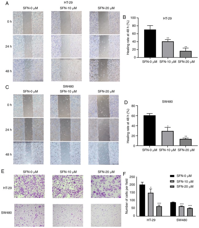
Figure 3.
SFN intervention decreases wound healing rates and cell migration. (A and C) HT-29 and SW480 cells were treated under different conditions, and their cell migratory ability was assessed by wound healing assay (magnification, ×40). (B and D) The wound healing rates of various groups presented in the graphs of parts A and C were quantified. The statistical significance of the results was analyzed by two-way ANOVA. (E) A total of 10×104 HT-29 or SW480 cells in 200 µl serum-free Dulbecco's modified Eagle's medium with different concentrations of SFN were seeded on the upper chamber, and cell migration was assessed by Transwell assay. Representative images are presented (magnification, ×100 and ×200). (F) The number of cells per field of various groups of part E was quantified. The statistical significance of the results was analyzed by two-way ANOVA. The results are presented as the mean ± standard deviation of 3 independent experiments. *P<0.05, **P<0.01 and ***P<0.001. SFN, sulforaphane; ANOVA, analysis of variance.

