Figure 5.
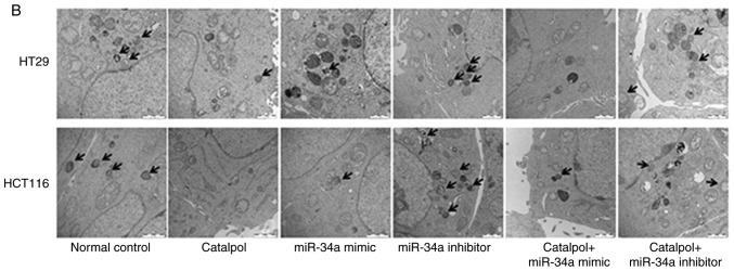
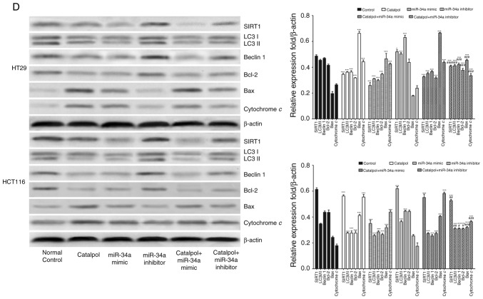
The miR-34a/SIRT1 axis participates in regulating catalpol-suppressed CRC cell autophagy and induces apoptosis. (A) HT29 and HCT116 cells were treated with 30 µM catalpol for 24 h and in combination with miR-34a mimic or inhibitor, and then flow cytometry was used to examine autophagic levels (***P<0.001 and ###P<0.001). (B) Autophagic vacuole formation was revealed by representative electron micrographs in CRC cell lines. The arrows indicate the autophagosomes. (C) The CRC cell apoptotic rate was analyzed by flow cytometry (***P<0.001 and ###P<0.001). (D) Western blotting revealed the expression of SIRT1, LC3, Beclin 1, Bcl-2, Bax, and cytochrome c in HT29 and HCT116 cells (*P<0.05 and ***P<0.001 indicates a significant difference vs. the normal control group; #P<0.05, ##P<0.01 and ###P<0.001 indicates a significant difference vs. the group treated with catalpol). miR, microRNA; SIRT1, sirtuin 1; CRC, colorectal cancer.


