Figure 2.
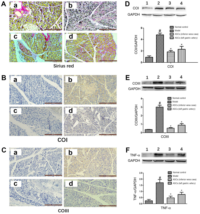
ASCs reduce pancreatic tissue collagen accumulation in DBTC-induced pancreatic fibrosis. (A) Pancreatic collagen was observed by Sirius Red staining (magnification, ×200). Using immunohistochemical staining (magnification, ×200) the expression of (B) COI and (C) COIII was detected. Images represent: a, the control; b, the DBTC-induced chronic pancreatitis model; c, the ASC-treated group I; and d, the ASC-treated group II. Using western blotting the expression of (D) COI, (E) COIII and (F) TNF-α were detected. Bands represent: 1, the control; 2, the DBTC-induced chronic pancreatitis model; 3, the ASC-treated group I; and 4, the ASC-treated group II. Data are presented as mean ± SEM (n=3). #P<0.05 vs. Control group; *P<0.05 vs. DBTC-induced chronic pancreatitis model group. ASC, adipose-derived mesenchymal stem cell; DBTC, dibutyltin dichloride; COI, collagen type I; COIII, collagen type III; TNF-α, tumor necrosis factor-α.

