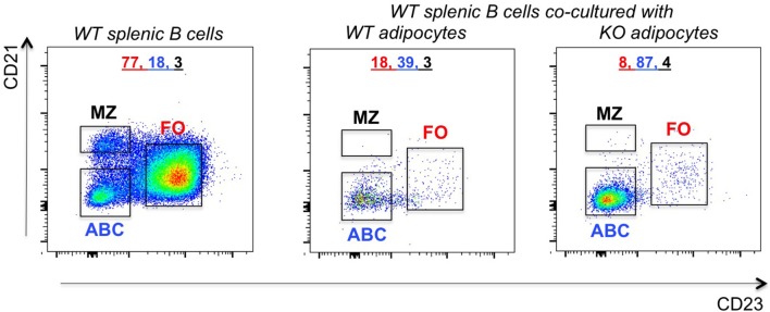
Figure 7.
Increased frequencies of ABCs in the VAT of P2KO vs. WT mice. B cells from the spleen of old WT mice were stained as in Figure 4 and frequencies of FO, ABC, MZ were evaluated. Adipocytes were isolated from the VAT of WT and P2KO old mice and cultured for 72 h in transwells with the splenic B cells of old WT mice. After this time, B cells were stained as indicated above and the frequencies of B cell subsets measured by flow cytometry. Results are representative of four independent experiments.

