
Figure 1.
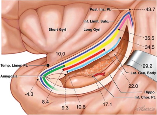
Anatomy of the insula. A schematic depiction of the insular region as would be viewed with retraction placed on the medial aspect of the superior temporal gyrus and incision through the inferior limiting sulcus of the insula. The insula is delimited anteriorly, superiorly, and inferiorly by the anterior, superior, and inferior limiting sulci of the insula, respectively. The insular cortex is arranged into anterior short and posterior long gyri. Fiber tracts coursing beneath the inferior limiting sulcus of the insula are represented. From superficial to deep: Extreme capsule (dark blue); uncinate fasciculus (green), inferior fronto-occipital fasciculus (yellow), and claustrocortical fibers (pink); anterior commissure (red); optic radiations (light blue). Distances of white matter pathways, the lateral geniculate body, and choroidal point from the limen insula are indicated. Modified with permission from Ribas et al., 2015