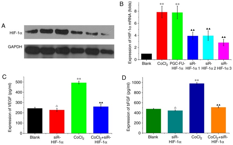
Figure 2.
Silencing of HIF-1α decreases VEGF and bFGF expression under hypoxic conditions in HCT-116 cells. (A) Western blotting of HIF-1α levels in HCT-116 cells. b-actin was used as the loading control. Three HIF-1α interference plasmids were used. Hypoxia was simulated in cells using 200 µmol/l CoCl2. (B) HIF-1α mRNA expression levels were analyzed in HCT-116 cells. The results revealed the fold change in the expression of HIF-1α normalized to GAPDH. (C) VEGF expression and (D) bFGF expression were analyzed in the supernatant of HCT-116 cells using ELISA under normal and hypoxic conditions. ΔP>0.05 vs. the normal control; **P<0.01 vs. the normal control; ▲▲P<0.01 vs. the hypoxic control. HIF-1α, hypoxia inducible factor-1α; VEGF, vascular endothelial growth factor; bFGF, basic fibroblast growth factor; CoCl2, cobalt chloride.

