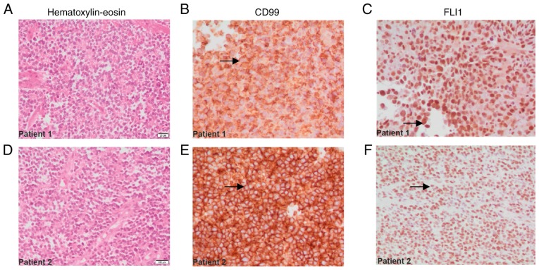
Figure 1.
Immunohistochemical analysis of lung tissue biopsy sections from patients with Ewing sarcoma. The samples of patient 1 are seen in A, B and C, and those of patient 2 are seen in D, E and F. (A and D) Small round cells, characteristic of tumors of the Ewing family, with hyperchromatic nuclei and small cytoplasmic space, as observed by hematoxylin and eosin staining. (B and E) Membranous staining for the CD99 protein (arrows). (C and F) Positive staining for the FLI1 transcription factor located in the nucleus (arrows). Magnification, ×40.

