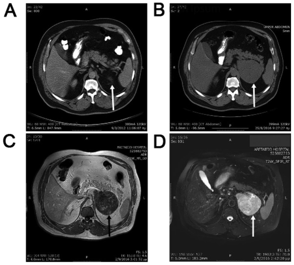
Figure 1.
(A) CT scan of the patient 4 years before diagnosis showing no pathology in the area of the left adrenal (arrow). (B) CT with intravenous contrast showing a 10.3x8.5x8.4 cm mass originating from the left adrenal gland (arrow), with minor contrast uptake. (C) MRI with low signal intensity on T1-weighted images (arrow). (D) MRI with high signal intensity on T2-weighted images (arrow). CT, computed tomography; MRI, magnetic resonance imaging.

