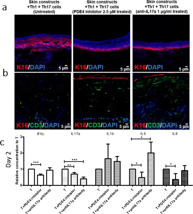
Figure 5.
Effect of PDE4 inhibitor and anti-IL17a antibody on Th1/Th17-incorporated 3D skin. Immunofluorescence staining of (a) K16 (red) and (b) CD3 (green) in Th1/Th17-bearing HSCs without treatment (left panel), with 2.5 μM PDE4 inhibitor treatment (middle panel), and 1 μg/ml anti-IL17a antibody treatment (left panel). (c) Relative cytokine secretion of IFNγ, IL-17a, IL-1b, IL-6, and IL-8 from Th1/Th17-bearing HSCs after PDE4 inhibitor or anti-IL17a antibody treatment on day 2.

