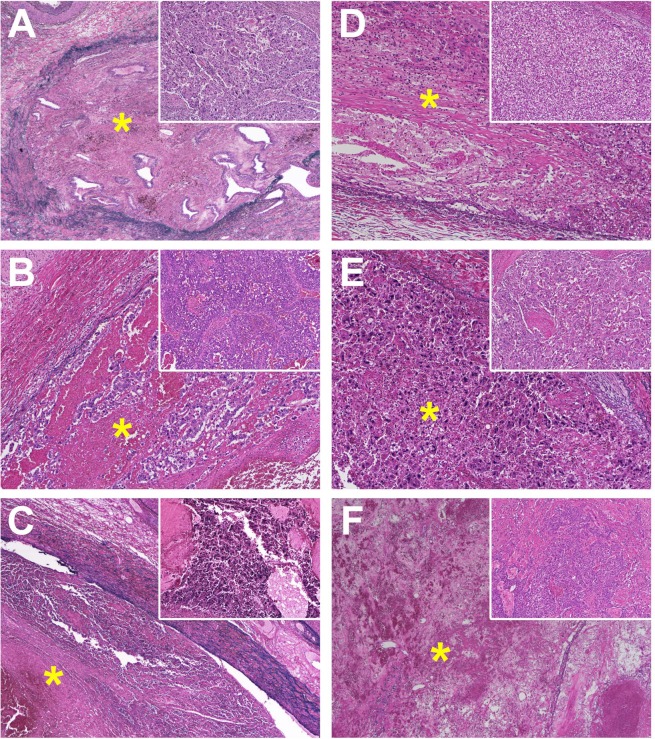
Figure 1.
Photomicrograph of thrombi in the portal vein (*; Victoria blue and haematoxylin-eosin stain [VB-HE]) with the primary tumour features (small windows in the right upper corner; VB-HE, x 50) in hepatocellular carcinomas surgically removed in patients (A to F). Portal-vein thrombi showing no evidence of viable tumour in Patients (A,C,F) (VB-HE, x 40), or containing residues of tumour cells in patients (B,D,E) with varying degrees of degeneration (VB-HE, x 100).

