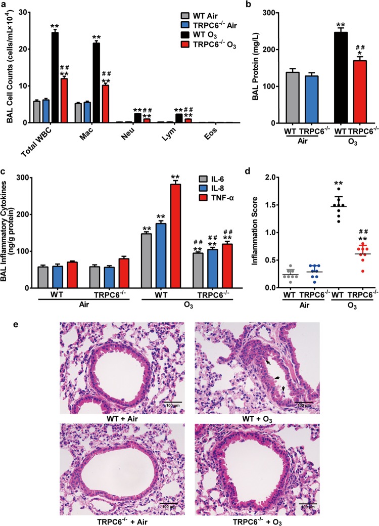
Fig. 1. Effect of TRPC6-deficiency on O3-induced airway inflammation.
WT and TRPC6−/− mice were exposed to O3 (1 ppm) for 3 h every other day (day 1, 3, 5).The mice were anesthetized 24 h after the last exposure. a–c Total white blood cell counts (Total WBC), macrophage counts (Mac), neutrophil counts (Neu), lymphocyte counts (Lym), eosinophil counts (Eos) (a), total protein content (b) and the release of inflammatory mediators IL-6, IL-8, TNF-α (c) in BAL fluid of different groups were compared. d Inflammation scores in air control and O3-exposed mice. e Representative histological sections of mouse lungs (H&E staining) after exposure to O3 or air. Black arrows: inflammatory changes. Scale bar: 100 μm. Results are presented as mean ± SEM, n = 8. *P < 0.05 or **P < 0.01 compared with WT + Air group, ##P < 0.01 compared with WT + O3 group.

