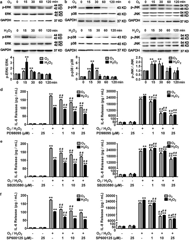
Fig. 7. Role of MAPK signal pathway in oxidative stress-induced inflammatory response in bronchial epithelial cells.
a–c Western blot analysis of phosphorylation protein expression of ERK (a), p38 (b) and JNK (c) after 16HBE cells were stimulated with O3 (100 ppb) or H2O2 (100 μM) for 0, 15, 30, 60, 120 min. **P < 0.01 compared with 0-min group. d–f After pretreatment with or without PD98059 (1, 10, 25 μM) (d), SB203580 (1, 10, 25 μM) (e) or SP600125 (1, 10, 25 μM) (f) for 1 h, 16HBE cells were stimulated with O3 (100 ppb) for 6 h followed by continuing culture for another 24 h or stimulated with H2O2 (100 μM) for 24 h. Release levels of IL-6 and IL-8 were detected. Data represent the mean ± SEM, n = 5. **P < 0.01 compared with Control group, ##P < 0.01 compared with H2O2 or O3 group.

