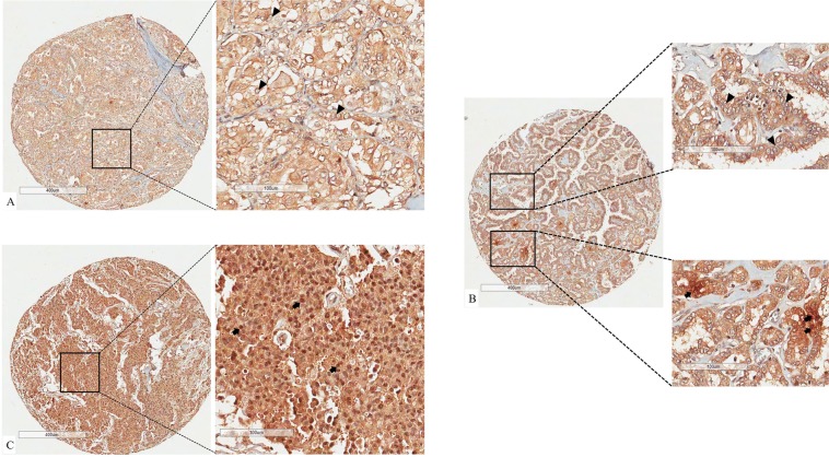
Figure 1.
Different percentages of nuclear RORγt positivity. Panel (A) shows the diffuse brownish in cytoplasm but not in nuclear of thyroid carcinoma cells. Panel (B) evidence that major of nuclei of thyroid cancer cells are negative for RORγt expression (upper right panel). Lower right panel shows a detail of focus of nuclei positive for RORγt. Panel C represent a tissue spot in which all nuclei of thyroid cancer cells are positive for RORγt. Black arrows point to nuclei positive for RORγt. Black arrow head point to nucleai negative for RORγt. Stromal components like collagen, vessels and myocytes are also not expected to express nuclear RORγt, giving us a negative control for each spot analyzed.

