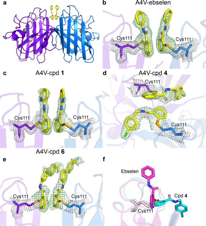
Fig. 3. The structures of A4V SOD1 with bound ligands.
a Cartoon representation of A4V SOD1 dimer with ligands. The ligands (yellow sticks with valences) bind each monomer (purple and blue) at Cys111, located at the dimer interface. Dark grey spheres represent Zn in the Zn binding sites and light grey spheres represent Zn in the Cu binding site. Details of ligand binding of b A4V-ebselen, c A4V-compound 1, d A4V-compound 4 and e A4V-compound 6. The Fo-Fc map contoured at 3σ (green) shows the electron density of the ligands. The 2Fo-Fc map contoured at 1σ (grey) shows the electron density of Cys111 residues. f The superposed structures of A4V-ebselen (magenta) and A4V-compound 4 (cyan) monomers, depicting the binding pose of each compound.

