
Figure 1.
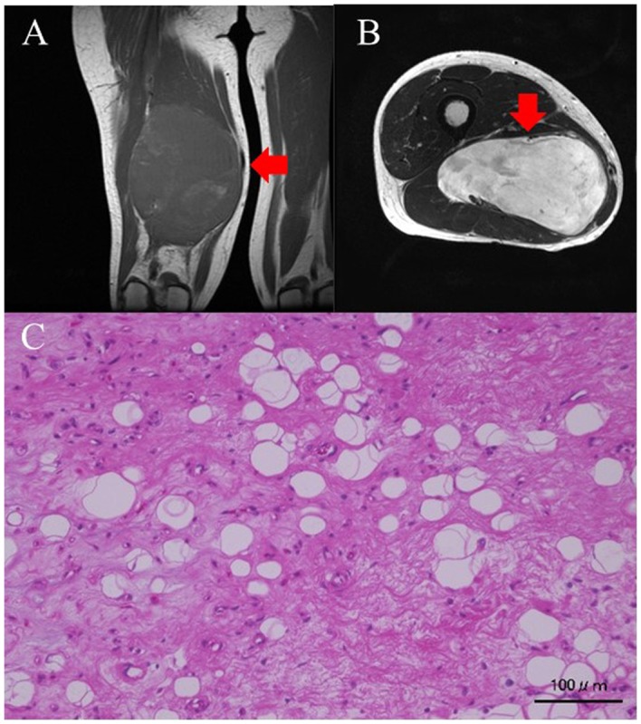
(A) Coronal T1-weighed MRI image of the thigh. The tumor intensity is low. (B) Transverse slices of T2-weighed MRI images of the thigh. The tumor intensity is high. (C) Histological findings on hematoxylin-eosin (H&E) staining. Small atypical cells proliferating in the background of a mucus matrix. Some lipoblasts are also noted. Red arrows indicate the tumor mass. Scale bar, 100 µm.