Figure 4.
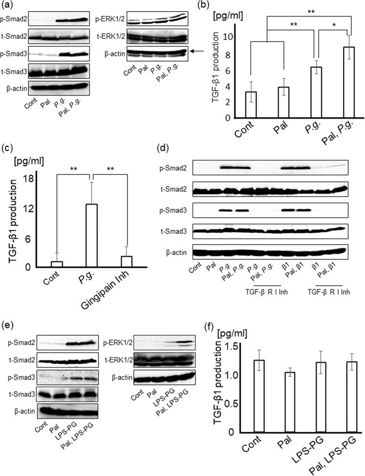
P.g.-infection and LPS-PG induce myofibroblastic activation of HSCs through Smad and ERK signaling pathways. P.g. infection induces TGF-β1 production through PAR2 activation by gingipain. (a) Smad2, Smad3, and ERK1/2 were detected by western blotting at 24 hours after P.g.-infection. (b) LX-2 cells with/without palmitate treatment were cultured with P.g (MOI-100) for 24 hours. The amount of TGF-β1 in each supernatant was measured by ELISA. Cont/Pal: N = 5, P.g./Pal, P.g.: N = 6. (c) LX-2 cells with/without gingipain inhibitors (3 µM) were cultured with P.g. (MOI-100) for 24 hours. Cont/Gingipain Inh: N = 8, P.g.: N = 7. (d) After culturing for 24 hours with/without TGF-β receptor I inhibitor (1 μg/ml), LX-2 cells were cultured with P.g. (MOI-100) for 24 hours. Smad2 and Smad3 were detected by western blotting. (e) Smad2, Smad3, and ERK1/2 were detected by western blotting at 4 days after LPS-PG stimulation. (f) LX-2 cells were cultured with LPS-PG (1 μg/ml) for 24 hours. The amount of TGF-β1 in each supernatant was measured by ELISA. Cont/Pal/LPS-PG/Pal, LPS-PG: N = 5. β-actin was used as internal control. Results were shown as mean ± SD. *p < 0.05, **p < 0.01. Pal: palmitate, P.g.: P.g.-infection, Gingipain Inh: Gingipain inhibitor, β1: TGF-β1, TGF-β R I Inh: TGF-β receptor I inhibitor.

