
Figure 2: C. parvum infection of human intestinal enteroid (HIE) monolayers in 2D human intestinal tissue model.
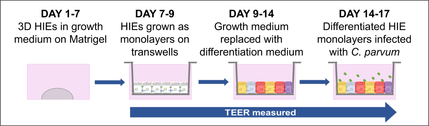
HIEs are grown in Matrigel in 24-well plates in growth medium for 7 days. HIEs are dissociated into single cells and grown as a monolayer on transwell inserts in growth medium for 3 days after which the growth medium is changed to differentiation medium for 5 days and the apical surface infected with C. parvum. Transepithelial electrical resistance (TEER) is measured from Days 7 to 17. Figure modified and reproduced with permission from Springer Nature Methods in Molecular Biology DOI 10.1007/7651_2017_1 © Springer Science+Business Media New York 2017. Winnie Y. Zou, et al Human Intestinal Enteroids: New Models to Study Gastrointestinal Virus Infections [21]