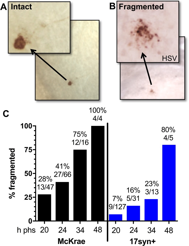
Fig 2. Neuronal fragmentation over time post-reactivation in vivo.
Latently infected mice were subjected to hyperthermic stress and ganglia were harvested at the indicated times post-hyperthermic stress (minimum of 10 ganglia per time point, see Fig 1C and 1D). Whole ganglia were processed for HSV protein expression. The brown precipitate (DAB) marks viral exit from latency. (A and B) Photomicrographs of portions of whole mount TG showing viral protein positive neurons that are (A) intact or (B) fragmented. Arrows depict higher magnification of neurons. (C) Neurons represented in Fig 1C and Fig 1D were evaluated with respect to the intact or fragmented phenotype. The percentage of fragmented neurons out of the total number of antigen positive neurons is indicated at each time post-hyperthermic stress examined. Fragmentation increased significantly over time post-reactivation in both McKrae (Chi square; p = 0.0008) and 17syn+ (Chi square; p<0.0001) infected TG.

