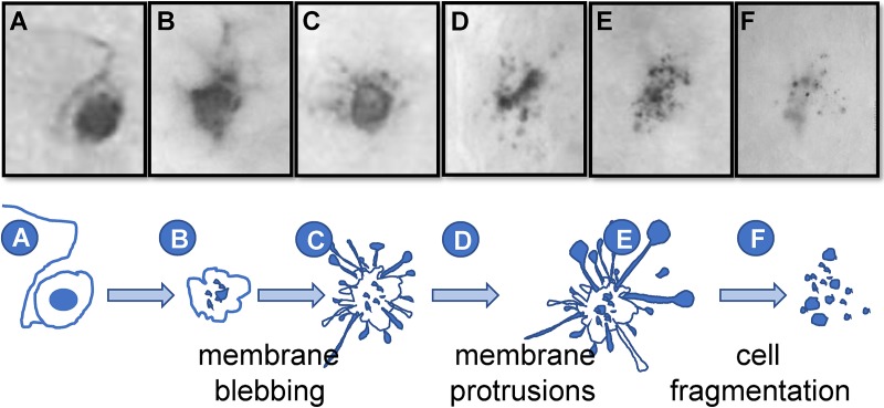
Fig 3. Proposed progression of neuronal destruction during reactivation in vivo.
Latently infected mice were subjected to hyperthermic stress and ganglia were harvested 20–48 h phs. Whole ganglia were processed for HSV protein expression. The dark precipitate (DAB) marks viral protein expression. (A-F) Based on the well-characterized progression of cells undergoing apoptosis, a schematic of the proposed progression from an intact to fragmented state of TG neurons undergoing HSV reactivation is shown. Photomicrographs of actual viral protein positive neurons that are: (A) intact, (B) early membrane blebbing, (C) prominent membrane protrusions, (D) fully fragmented with prominent apoptotic bodies, and (E and F) fully fragmented with a small cluster of apoptotic bodies remaining are shown above the schematic and correspond with the letters indicated at each stage on the schematic.

