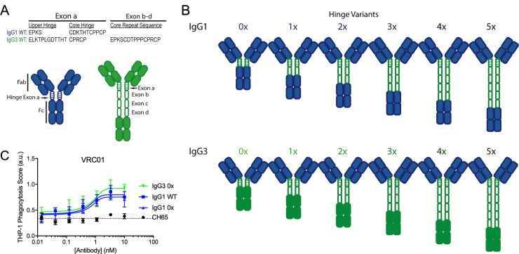
Fig 2. IgG1 and IgG3 hinge variant panel.
A. Sequences of natural IgG1 and IgG3 hinge exons, noting the upper and core hinge regions encoded by exon a and the core hinge repeat sequences encoded by exons b-d in IgG3. B. Schematic of the panel of hinge swapped and extended IgG1 and IgG3 variants. Domains derived from IgG1 and IgG3 are indicated in blue and green respectively. C. Phagocytic activity of VRC01 WT IgG1 and 0x forms of IgG1 and IgG3 in the THP-1 ADCP assay against CH505TF gp140 antigen-conjugated beads. Error bars indicate mean and SD of duplicates. Dotted horizontal line represent phagocytosis value observed in the absence of Ab. Phagocytic scores for CH65, an IgG1 Ab specific for hemagglutinin are shown as an additional negative control. Connecting lines indicate curve fit models. AU: arbitrary units.

