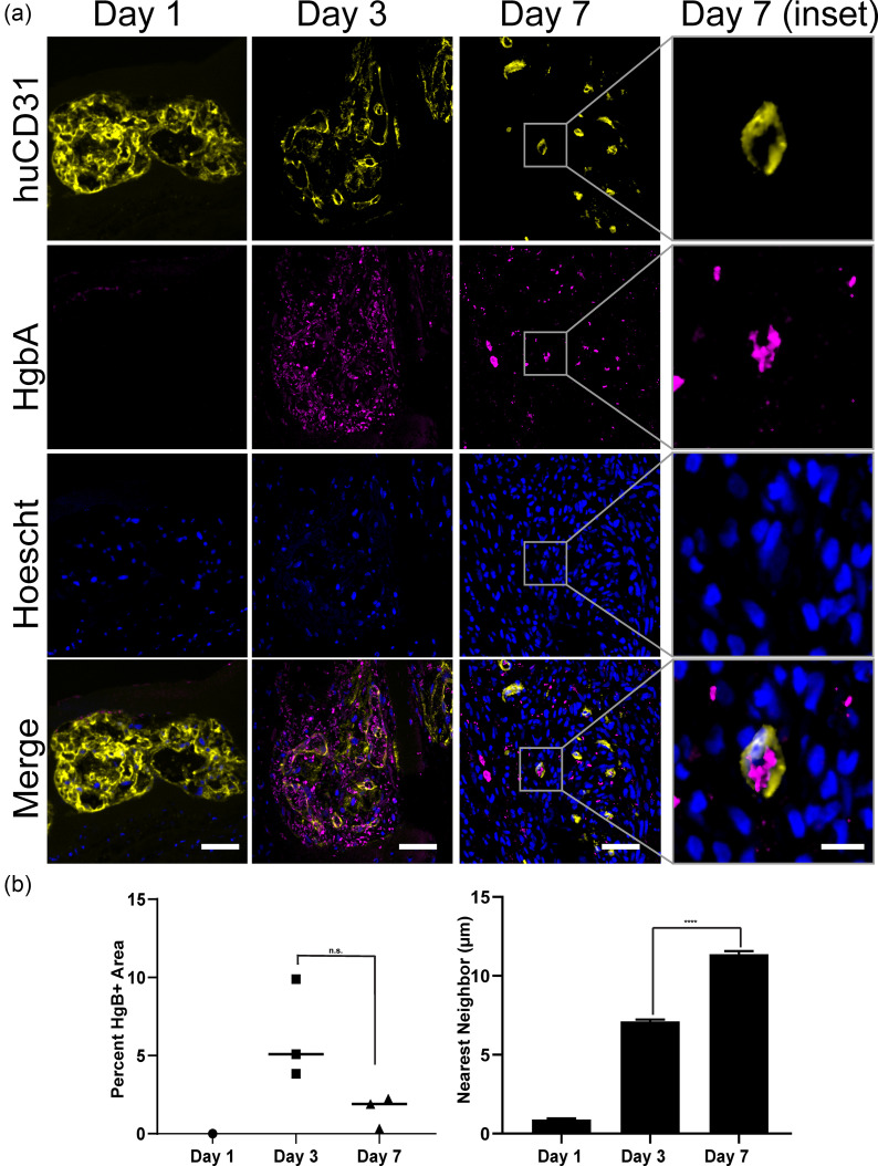
FIG. 4.
Graft-derived vessels are perfused with blood. (a) Co-staining for HgbA and huCD31 in explants from 1, 3, and 7 days. Scale bar = 50 μm. Inset scale bar = 10 μm. (b) Quantification of HgbA staining as the percent of patch area and distance to the nearest neighbor. (***p < 0.001). n = 1 for day 1 and n = 3 for day 3 and day 7. Error bars indicate S.E.M.

