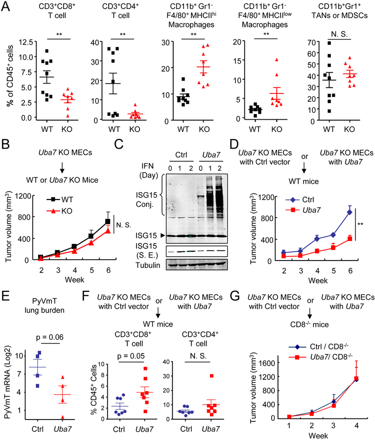
Figure 2. Protein ISGylation stimulates intratumoral infiltration of T lymphocytes.
A, Flow cytometric analysis of intratumoral leukocytes in the tumors from PyVmT/WT and PyVmT/Uba7 KO mice. Numbers expressed as percentages of total CD45+ leukocytes (n = 7–8). p, student’s t-test. TANs, tumor-associated neutrophils; MDSCs, myeloid-derived suppressor cells.
B, Tumor growth from PyVmT/Uba7 KO MECs in WT and Uba7 KO Mice (mean + SEM, n = 5). p, two-way ANOVA.
C, Restored Uba7 expression in PyVmT/Uba7 KO MECs. Cells were treated by type I IFN up to two days and cell lysate were immunoblotted by antibodies as indicated. S. E., short exposure.
D, Tumor growth from PyVmT/Uba7 KO + control vector (henceforth “Ctrl”) or PyVmT/Uba7 KO + Uba7 (henceforth “Uba7”) MECs in WT mice (mean + SEM, n = 4). A representative set from three independent experiments (n = 4–5) is shown. **, p<0.01, by two-way ANOVA.
E, PyVmT lung burden in mice from panel D (mean + SEM, n = 4). p, student’s t-test.
F, Flow cytometric analysis of CD4+ and CD8+ T cell in tumors from mice injected with Ctrl or Uba7 MECs after week 6. Numbers expressed as percentages of total CD45+ leukocytes. Pooled data are presented from two independent experiments (n = 7). p, student’s t-test.
G, Tumor growth from Ctrl or Uba7 MECs in CD8 deficient mice (mean + SEM, n = 6).

