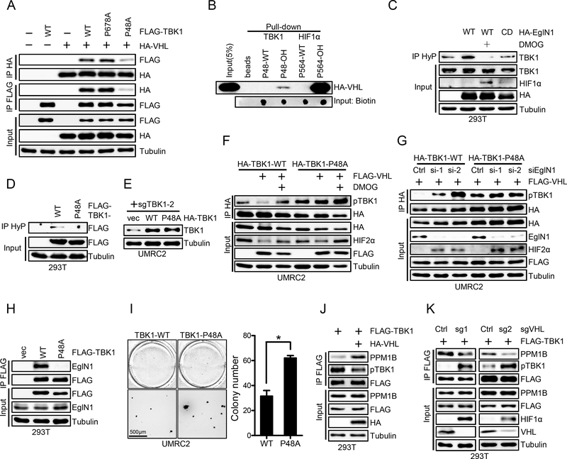
Figure 2. Hydroxylated Pro48 mediates TBK1-VHL interaction.
A, Immunoblots of whole cell lysates (input) and immunoprecipitations (IP) from 293T cells transfected with indicated plasmids.
B, Immunoblots of peptide pull-down samples as indicated.
C-D, Immunoblots of whole cell lysates (input) and immunoprecipitations (IP) from 293T cells transfected with indicated plasmid and treated with DMOG (1mM) as indicated. HyP denotes pan-hydroxyPro antibody.
E, Immunoblots of lysates from UMRC2 cells infected with lentivirus encoding vec, HA-TBK1 or HA-TBK1-P48A and then infected with lentivirus encoding TBK1 sgRNA(sg2).
F-G, Immunoblots of whole cell lysates (input) and immunoprecipitations (IP) from UMRC2 cells infected with lentivirus encoding HA-TBK1-WT or HA-TBK1-P48A and transfected with indicated plasmids or siRNA, followed by indicated treatment.
H, Immunoblots of whole cell lysates (input) and immunoprecipitations (IP) from 293T cells transfected with indicated plasmids.
I, Representative soft agar growth and quantification of colony number (duplicate wells) of UMRC2 cells infected with lentivirus encoding HA-TBK1 or HA-TBK1-P48A and then transfected with HA-VHL. See Fig. 2E for HA-TBK1 expression. Error bars represent SEM, * P<0.05.
J-K, Immunoblots of whole cell lysates (Input) and immunoprecipitations (IP) from 293T cells infected with indicated lentivirus and transfected with indicated plasmids.

