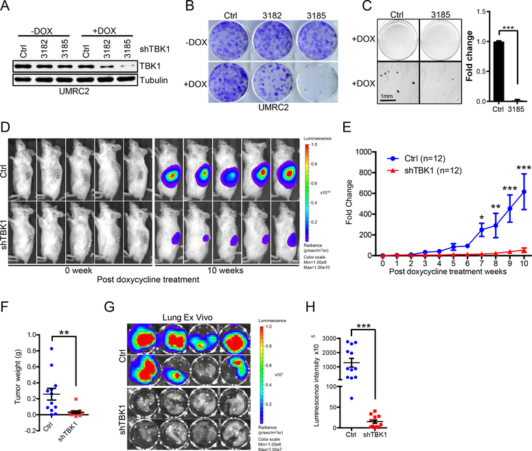
Figure 4. Loss of TBK1 selectively suppresses VHL null ccRCC tumor growth.
A-C, Immunoblots of lysates (A) from, representative crystal violet staining (B) and 3-D soft agar growth pictures (C) of UMRC2 cells infected with lentivirus encoding either inducible Ctrl shRNA or inducible TBK1 shRNA and then treated with doxycycline as indicated.
D-E, Representative bioluminescence imaging of before (0 week) and 10 weeks post-doxycycline treatment (D) and quantification of post-doxycycline treatment bioluminescence imaging (E) from UMRC2 luciferase stable cells infected with lentivirus encoding either Teton-control (Ctrl) Teton-shTBK1 3185 (shTBK1) injected orthotopically into the renal capsule of NSG mice. Two-way ANOVA analysis was performed for E.
F, Quantification of kidney tumor weight (n=12).
G-H, Representative lung ex vivo bioluminescence imaging (G) and quantification of ex vivo imaging (H, n=12).
Error bars represent SEM, *P<0.05, **P<0.01, ***P<0.001

