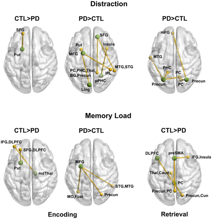
Figure 4.

Group differences in context‐dependent functional connectivity during working memory. The figure illustrates significant group differences in the connectivity of a seed ROI (green circles) with other brain regions (gold circles and lines) during the encoding and retrieval phases of the task. Top row: Group differences in distraction‐dependent connectivity. Bottom row: Group differences in memory load‐dependent connectivity. Tables 2 and 3 provide the characteristics of distraction and load‐related connectivity features that differed between groups. BG, basal ganglia; DLPFC, dorsal lateral prefrontal cortex; Fusi, fusiform gyrus; IFG, inferior frontal gyrus; IPL, inferior parietal lobule; mdThal, mediodorsal thalamus; mFG, medial frontal gyrus (BA 10); MFG, middle frontal gyrus (BA 6); MO, middle occipital (BA 19); MTG, middle temporal gyrus; PC, posterior cingulate; precun, precuneus; PHC, parahippocampal cortex; pPHC, posterior parahippocampal cortex; SFG, superior frontal gyrus (BA 8); preSMA, presupplementary motor area; STG, superior temporal gyrus; Thal, thalamus
