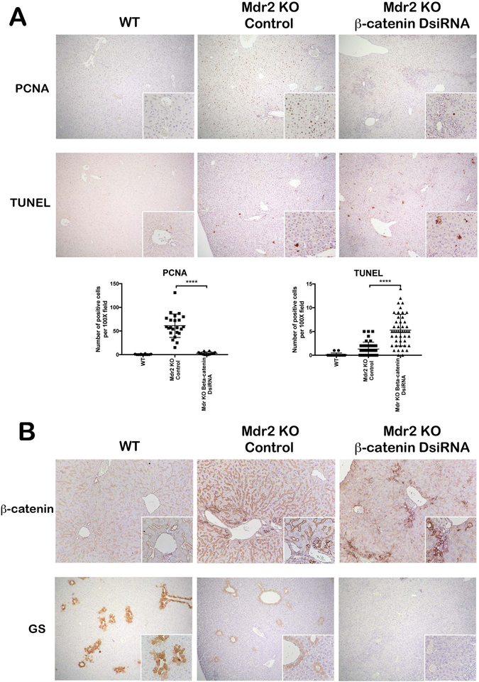
Figure 3: Simultaneous loss of Mdr2 and β-catenin leads to impaired regenerative response in liver.
IHC for PCNA shows increased proliferative response in Mdr2 KO mice, which was significantly reduced in KO/KD liver (50X; 100X inset). TUNEL staining revealed increased cell death in KO/KD liver compared to Mdr2 KO alone (50X; 100X inset). Quantification of PCNA and TUNEL staining further confirms the IHC interpretation. (B) IHC for β-catenin showed significantly reduced expression in KO/KD hepatocytes compared to WT or KO alone (100X; inset 200X). GS staining is reduced in Mdr2 KO as compared to WT and absent in KO/KD liver (50X; 100X inset). ****p<0.0001.

