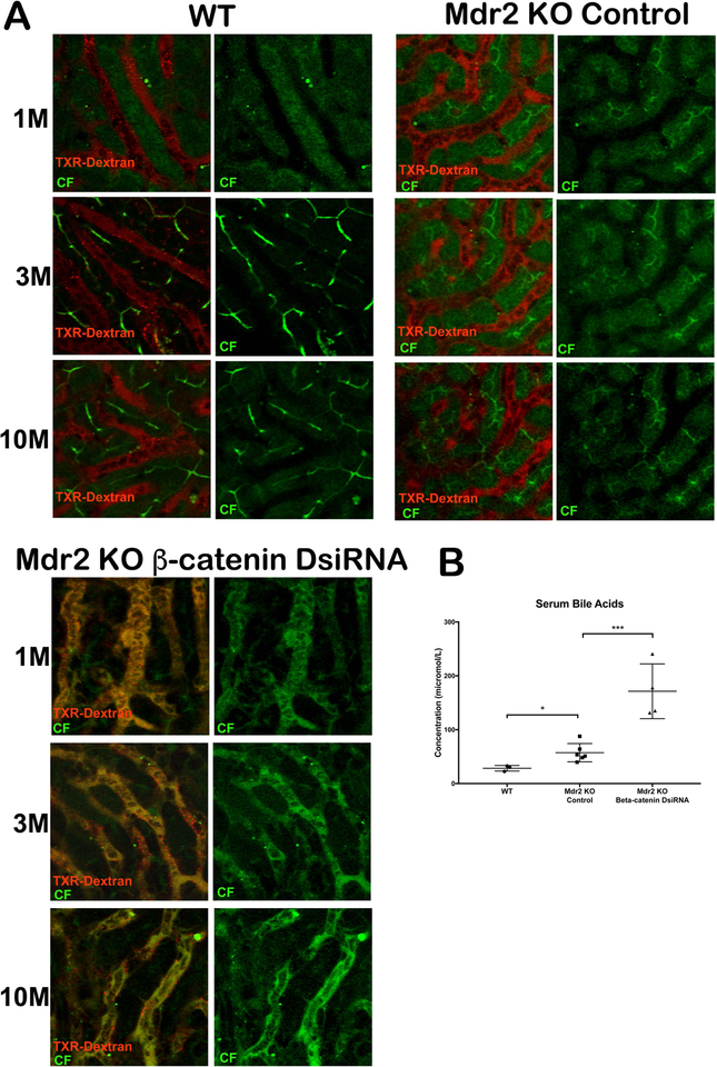
Figure 7: Dual loss of β-catenin and Mdr2 in hepatocytes leads to extrusion of bile into hepatic sinusoids.
(A)Time series images (1m, 3m and 10m) of WT liver shows localization of CF (green) and Texas-red dextran (red) in bile canaliculi and sinusoids, respectively. Mdr2 KO alone showed similar localization of CF and TXR-dextran. However, KO/KD liver shows co-localization of CF and Texas-red dextran in sinusoids and failure of secretion into bile canaliculi at all time points after injection of dyes. Scale bars: 50μm. (B) Levels of serum BA are significantly higher in KO/KD compared to KO alone *p<0.05; ***p<0.001.

