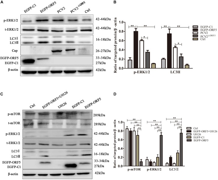
FIGURE 4.
ORF5 protein upregulates phosphorylation of ERK1/2. (A) EGFP-ORF5 was transfected for 48 h, PCV2 or PCV2ΔORF5 was infected for 36 h, p-ERK1/2, t-ERK1/2, LC3II were detected by immunoblotting. (B) Perform a T-test on the grayscale analysis of the A picture, the ratio of targeted protein to β-actin. (C) PK-15 cells were pretreated with U0126 for 2 h and then transfected with EGFP-ORF5 or EGFP-C1 for 48 h, p-mTOR, t-mTOR, p-ERK1/2, t-ERK1/2, LC3II were detected by immunoblotting. (D) Perform a T-test on the grayscale analysis of the (C), the ratio of targeted protein to β-actin. *p < 0.05, **p < 0.01.

