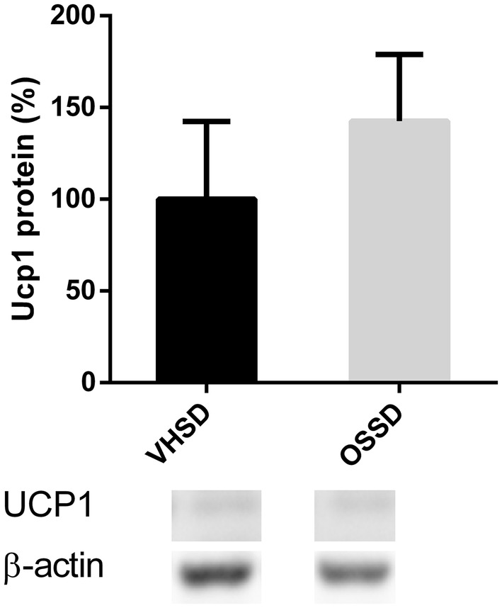
Fig. 2.

UCP1 protein levels in BAT measured by Western blotting of Fischer 344 rats supplemented with orange lyophilizate from the southern hemisphere (OS) or vehicle (VH) held in a short-day photoperiod (SD). Data are normalized to β-actin and to the VHSD group, set at 100%. Results are presented as the mean ± SEM and data compared with Student’s t test (p < 0.05)
