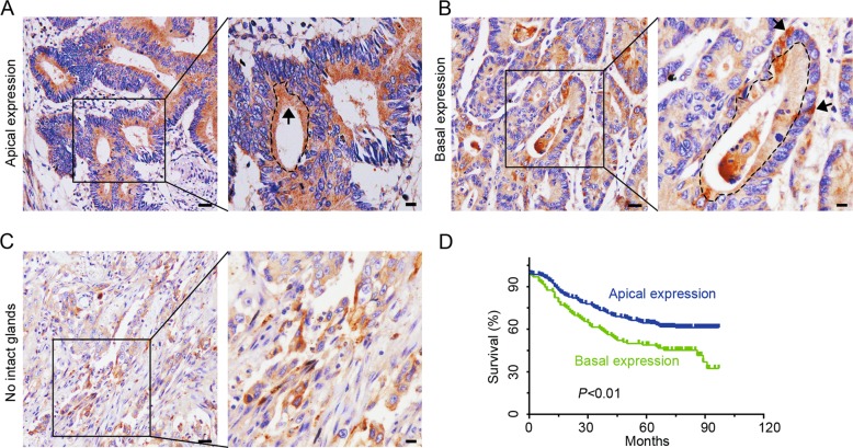
Fig. 1. Cdc42 expression patterns as determined by immunohistochemical staining of CRC tissue.
a, b CRC tissue with apical or basal expression. Arrows indicate the Cdc42 distribution, and the dotted curve represents the glandular cavity. Scale bars represent 50 or 10 μm. c Samples lacked intact glands in poorly differentiated adenocarcinoma tissue sections (scale bars: 50 μm, bars in enlarged images: 10 μm). d A Kaplan–Meier survival curve for CRC patients with Cdc42 expression in their tumour cells.

