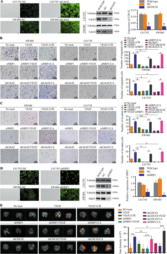
Fig. 6. Activated Cdc42 promotes migration/invasion and lung metastases of CRC cells.
a LS174T and SW480 cells were transfected with NC or shCdc42 in the first column, scale bar, 100 μm, and Cdc42 expression was detected using western blotting in the second column. Cdc42 was amplified using real-time quantitative polymerase chain reaction in the third column. NC represents cells infected with empty lentiviruses and was used as a negative control. Error bars represent the mean ± SD of triplicate experiments; *P < 0.001. b Transwell migration activity of SW480 and LS174T cells induced by different conditions and representative images. Data are the means ± SD from three independent experiments, *P < 0.001. Scale bar, 50 μm. c Representative images of the Transwell invasion assay showed that SW480 and LS174T cells were induced by different conditioned media. Data are presented as the means ± SD from three independent experiments, *P < 0.01, **P < 0.001. Scale bar, 50 μm. d LS174T and SW480 cells were transfected with NC or shNRP1 in the first column, scale bar, 100 μm, and NRP1 expression was detected using western blotting in the second column. NRP1 was amplified using real-time quantitative polymerase chain reaction in the third column. NC represents the cells infected with empty lentivirus and was used as a negative control. Error bars represent the mean ± SD of triplicate experiments; *P < 0.001. e Representative bioluminescent images of nude mouse lungs 30 days after intravenous injection. f Quantification analysis of fluorescence signals from captured bioluminescence images. *P < 0.05, **P < 0.01 and ***P < 0.001.

