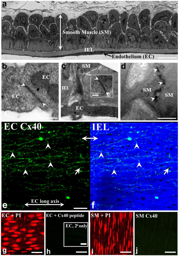
Figure 1: Vascular cells in cerebral arteries connect to one another and express Cx40.
(a) Electron microscopy reveals that mouse middle cerebral arteries are composed of smooth muscle (SM), internal elastic lamina (IEL) and endothelial cell (EC) layers. Bar = 5 μm. (b-d) Contact sites (arrowheads) between endothelial cells (b, bar = 0.25 μm), between smooth muscle and endothelium (c, scale bar 0.25 μm), and between smooth muscle cells (d, bar = 0.5 μm). (e) Cx40 (green), a key gap junctional protein was identified by immunohistochemistry in middle cerebral arteries isolated from mice. (f) IEL was delineated by 488 nm autofluorescence (g,i) Propidium iodide labeling verified EC and SMC patency. (h) Endothelial Cx40 signal was absent in sections incubated without the primary antibody (inset) and in endothelial cells treated with a Cx40 peptide that competes for the primary antibody binding site. (j) Cx40 staining was also absent in the smooth muscle cell layer. Bar, e-j = 25 μm

